LONG CASE
51 year old male patient who is resident of chityal ,and works in a transportation company came to the hospital with complaints of
1- Fever since 10 days
2- Cough since 10 days
3-shortness of breath since 6 days
History of presenting illness :
Patient was apparently asymptomatic 10 days back, then he developed....
Fever since 10 days which was high grade , with chills and rigors , Intermittent, relievedwith medication.
Associated with cough and shortness of breath.
Cough since 10 days which is productive ,mucoid in consistency,whitish ,scanty amount ,more during night times and on supine position ,non foulsmelling ,non bloodstained .
Right sided chest pain - diffuse , intermittent ,dragging type , aggravated on cough ,non radiating ,not associated with sweating , palpitations.
Shortness of breath since 6 days , insidious onset , gradually progresive ,of grade 3 - (MMRCscale) ,not associated with wheeze ,no orthopnea ,no Paroxysmal nocturnal dyspnea, no pedal edema .
History of pain abdomen or abdominal distension.
No history of , vomiting ,loose stools .
No history of burning micturition.
Past history :
Patient gives history jaundice 15 days back that resolved in a week .
No history of Diabetes , Hypertension , Tuberculosis ,Bronchial asthma ,COPD , coronary artery disease , Cerebrovascular accident ,thyroid disease.
Family history :
No history of Tuberculosis or similar illness in the family
Personal history :
Patient is a chronic smoker - smokes 5 cigarettes per day from past 25 years .
He is a Chronic alcoholic - cosumes 300 ml whisky per day ,but stopped since 3 months.
No bowel and bladder disturbances
Summary :
51 year old male patient with fever ,cough , shortness of breath possible differentials
1- Pneumonia
2- Pleural effusion
GENERAL EXAMINATION :
Patient is moderately built and nourished.
He is conscious, cooperative,comfortable.
No signs of pallor ,cyanosis ,icterus ,koilonychia , lymphadenopathy ,edema .
Vitals :
Patient is afebrile .
Pulse - 86 beats / min ,normal voulme ,regular rhythm,normal character ,no radiofemoral delay,radioradial delay.
BP - 110/70 mmhg ,measured in supine position in both arms .
Respiratory rate -22 breaths / min


SYSTEMIC EXAMINATION :
Patient examined in sitting position
Inspection:-
Upper respiratory tract - oral cavity- Nicotine staining seen on teeth and gums , nose & oropharynx appears normal.
Chest appears Bilaterally symmetrical & elliptical in shape
Respiratory movements appear to be decreased on right side and it's Abdominothoracic type.
Trachea is central in position & Nipples are in 4th Intercoastal space
Apex impulse visible in 5th intercostal space
No signs of volume loss
No dilated veins, scars, sinuses, visible pulsations.
No rib crowding ,no accessory muscle usage.
Palpation:-
All inspiratory findings are confirmed by palpation.
Spine position is normal and no tenderness seen.
Trachea central in position
Apical impulse in left 5th ICS, 1cm medial to mid clavicular line.
Cricosternal distance is 3finger breadths.
PERCUSSION:stony dullness is observed (large pleural effusion)
AUSCULTATION
Other systems examination :
Gastrointestinal system :
Inspection -
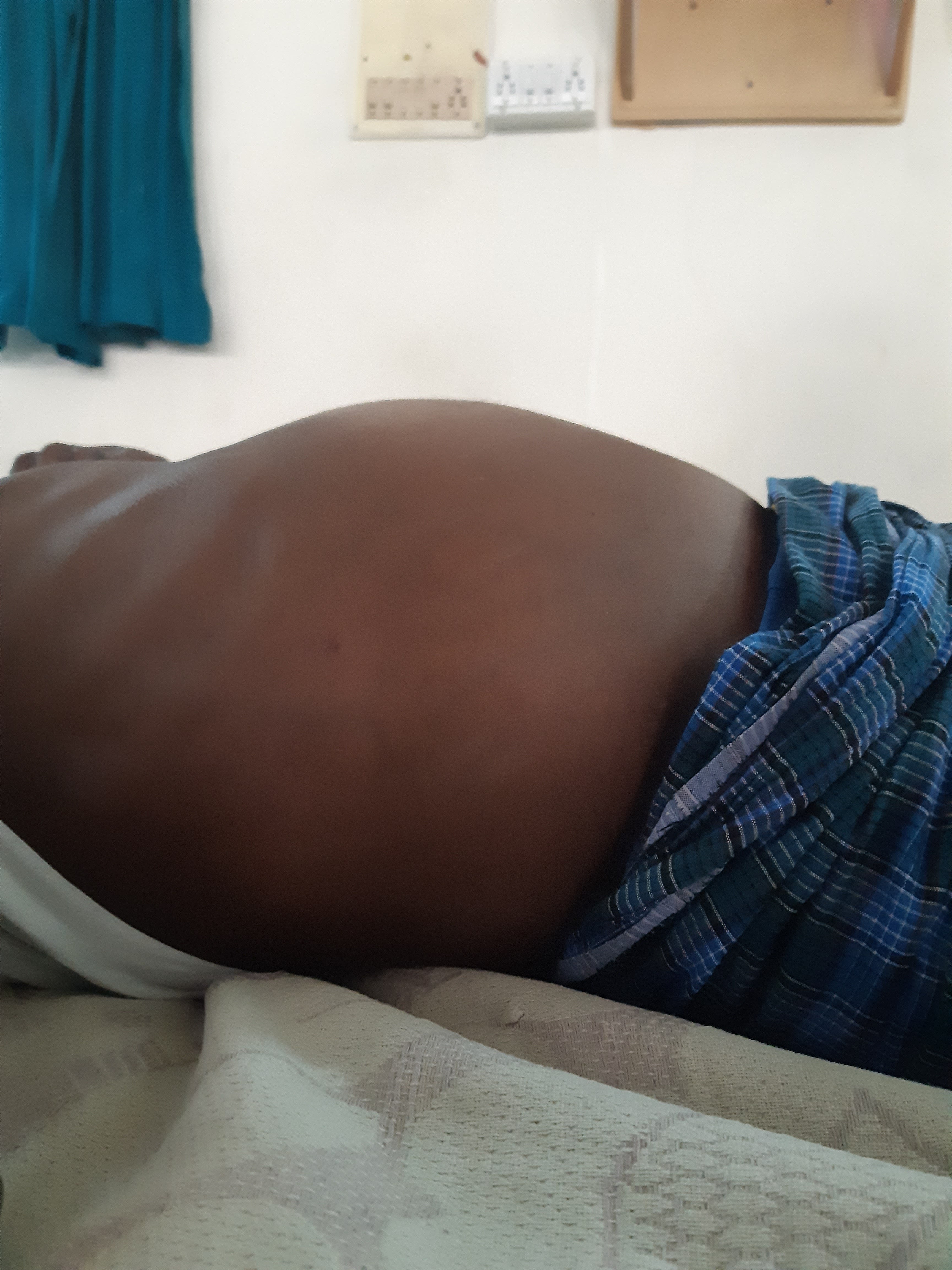
Abdomen is distended.
Umbilicus is central in position and slightly retracted and inverted.
All quadrants of abdomen are equally moving with respiration except Right upper quadrant .
No visibe sinuses ,scars , visible pulsations or visible peristalsis
Palpation and Percussion:
All inspectory findings are confirmed.
No tenderness on palpation.
Liver - is palpable 4 cm below the costal margin and moving with respiration.
Liver span increased(18cm)- normal is 13cm
Spleen : not palpable.
Kidneys - bimanually palpable
Percussion is normal.
Auscultation- bowel sounds heard .
No bruits and venous hum.
Cardiovascular system -
S1 and S 2 heard in all areas ,no murmurs
Central nervous system - Normal
Per rectal examination_ Normal
Final Diagnosis :
1- Right sided Pleural effusion likely infectious etiology.
2- Hepatomegaly - ? Hepatitis or ? Chronic liver disease
Investigations :
X ray findngs-ELLIS curve (s shaped curve/Damoiseaus curve)-curved shadow at the lung base,blunting the costophernic angle and ascending towards the axilla.
Shifting dullness is seen on examination
Pleural fluid analysis :
Colour - straw coloured
Total count -2250 cells
Differential count -60% Lymphocyte ,40% Neutrophils
No malignant cells.
Pleural fluid sugar = 128 mg/dl
Pleural fluid protein / serum protein= 5.1/7 = 0.7
Pleural fluid LDH / serum LDH = 190/240= 0.6
Interpretation: Exudative pleural effusion.
Other investigations :
Serology negative
Serum creatinine-0.8 mg/dl
Clinical urine tests -Normal
LIVER FUNCTION TESTS
CT abdomen :
Final Diagnosis:
1-Right sided Pleural effusion - synpneumonic effusion
2- Right lobe liver abscess(12×11 cm partially liquified)
TREATMENT
- Inj. Piptaz 2.25mg IV QID
- Tab AZITHRO 500mg OD
- Inj METROGYL 100ml TID
- Inj Neomol 1gm/IV
- Tab Dolo 650mg
- O2 Inhalation
- Neb Duolin 8th hrly
- IV Normal Saline
- Inj Optineuron
- Temp monitoring 4thrly
- Bp and SpO2 monitoring
- Inj Amikacin
-----------------------------------------------------------------------------------------------------------------------
SHORT CASE
50 year old male, farmer by occupation, resident of Pochampally, came to Medicine OPD with complaints of :
* Distended abdomen since 7 days
* Pain abdomen since 7 days
* Pedal edema since 5 days
* Breathlessness since 4 days.
HISTORY OF PRESENT ILLNESS:
The patient was apparently asymptomatic 6 months ago when he developed jaundice and was treated at a private practitioner.
Later he developed abdominal distension about 7 days ago - insidious in onset, gradually progressive to the present size - associated with
- Pain in epigastric and right hypocondrium - colicky type.
- Fever - high grade, not associated with chills and rigor, decreased on medication, No night sweats.
- Not associated with Nausea, vomiting, loose stools
There was pedal edema
- Gradually progressive
- Pitting type
- Bilateral
- Below knees
- Increases during the day - maximum at evening.
- No local rise of temperature and tenderness
- Grade 2
- Not relived on rest
He also complained of shortness of breath since 4 days - MRC grade 4
- Insidious in onset
- Gradually progressive
- Agrevated on eating and lying down ; No relieving factors
- No PND
- No cough/sputum/hemoptysis
- No chest pain
- No wheezing
Patient is a known alcoholic since 20 years. Ascites increased after his last drink on 29th May, 2022.
Daily Routine :
Wakes up at 5am and goes to field.
Comes home at 8am and has rice for breakfast. Returns to work at 9am.
1pm - lunch
2-6 pm - work
6pm - home
8pm - dinner
Alcohol- 2 times a week, 180 ml.
PAST HISTORY:
No history of similar complaints in the past
Medical history- not a known case of DM, HTN, TB, Epilepsy, Asthma, CAD
Surgical history - not significant
PERSONAL HISTORY:
- Diet - mixed
- Appetite- reduced since 7 days
- Sleep - disturbed
- Bowel - regular
- Bladder - oliguria since 2 days, no burning micturition, feeling of incomplete voiding.
- Allergies- none
- Addictions - Beedi - 8-10/day since 20 years ;
- Alcohol - Toddy - 1 bottle, 2 times a week, since 20 years;
- Whiskey-180 ml, 2 times a week, since 5 years.
- Last alcohol intake - 29th May, 2022.
FAMILY HISTORY:
Not significant
GENERAL EXAMINATION:
Patient is conscious, coherent and co-operative.
Examined in a well lit room.
Moderately built and nourished
Icterus - present (sclera)
Pedal edema - present - bilateral pitting type, grade 2 - https://youtube.com/shorts/Uwz_0gxzqUM?feature=share
No pallor, cyanosis, clubbing, lymphoedenopathy.
Vitals :
Temperature- febrile
Respiratory rate - 16cpm
Pulse rate - 101 bpm
BP - 120/80 mm Hg.
SYSTEMIC EXAMINATION:
CVS : S1 S2 heard, no murmurs
Respiratory system : normal vesicular breath sounds heard.
Abdominal examination:
INSPECTION :
Shape of abdomen- distended
- Umblicus - everted
- Movements of abdominal wall - moves with respiration
- Skin is smooth and shiny;
- No scars, sinuses, distended veins, striae.
PALPATION :
Local rise of temperature present.
Tenderness present - epigastrium.
Tense abdomen
Guarding present
Rigidity absent
Fluid thrill positive
Liver not palpable
Spleen not palpable
Kidneys not palpable
Lymph nodes not palpable
PERCUSSION:
Liver span : not detectable
Fluid thrill: felt
AUSCULTATION:
Bowel sounds: heard in the right iliac region
CNS EXAMINATION:
Conscious
Speech normal
No signs of meningeal irritation
Cranial nerves: normal
Sensory system: normal
Motor system: normal
Reflexes: Right. Left.
Biceps. ++. ++
Triceps. ++. ++
Supinator ++. ++
Knee. ++. ++
Ankle ++. ++
Gait: normal
INVESTIGATIONS
Serology:
HIV - negative
HCV - negative
HBsAg - negative
PROVISIONAL DIAGNOSIS:
Acute decompensated liver failure with ascites.
TREATMENT:
Syp. Lactose 15ml TID
Abdominal girth charting - 4th hourly
Fluid restrictriction less than 1L per day
Salt restriction less than 2 gms per day


Comments
Post a Comment