1801006044 CASE PRESENTATION
- Get link
- X
- Other Apps
LONG CASE
50 year old Male came to the medicine OPD with chief complaints of
- Difficulty in breathing since 5 days , and an episode of sob early in the morning prior to admission
- Decreased urine output since 5 days
- Swelling of lower limbs on and off since 1 year
HISTORY OF PRESENTING ILLNESS
Patient was apparently asymptomatic 1 year ago ,then He went to local hospital and was diagnosed with hypertension and is on Telmisartan 40mg since 1 year, then he developed bilateral pedal edema on and off in nature since 1 year from knee to ankle region, and is on conservative treatment.
5 days ago during night patient developed sob sudden in onset and gradually progressive class 3, associated with orthopnea.
associated with PND
urine output was narrow streamlined urine
history of intermittent fever not associated with chills and rigor
not associated with chest pain
not associated with sweating
no history of burning micturition
DAILY ROUTINE
Patient wakes up at 5:30 in the morning and does his household chores and goes to work for 5 hours and comes back at 1 pm to have lunch, and takes rest for the day. Patient have dinner at around 7:30 in evening and goes to sleep at 9pm.
PAST HISTORY
Known case of hypertension for 1 year
Not a known case of DM, asthma, epilepsy, thyroid disorders.
DRUG HISTORY
Is on Telmisartan 40 mg since 1yr
FAMILY HISTORY
No similar complaints in the past
PERSONAL HISTORY
Appetite Normal
Diet mixed
Sleep Adequate
Bowel and bladder Regular, Decreased micturition
Addictions
Smoking -beedi consumer (4 beedis per day so 6 pack years)
Alcohol -since 25 years 4 times monthly(whisky 90 ml each time)
GENERAL EXAMINATION
Patient is conscious, coherent, and cooperative
moderately built and moderately nourished
Pallor - present
Icterus-absent
Cyanosis - absent
Clubbing-absent
Lymphadenopathy -absent
Pedal edema -absent
vitals
Temperature - Afebrile
Pulse - 76 bpm
Blood pressure- 130/80 mmhg
Respiratory rate- 17 cycles per min
Spo2 - 95%
SYSTEMIC EXAMINATION
CVS :-
Inspection :
No palpitations
JVP seen
Palpation
Apex at 6th intercoastal space
No parasternal heave
No palpable P2
Auscultation
S1 S2 heard
RESPIRATORY SYSTEM
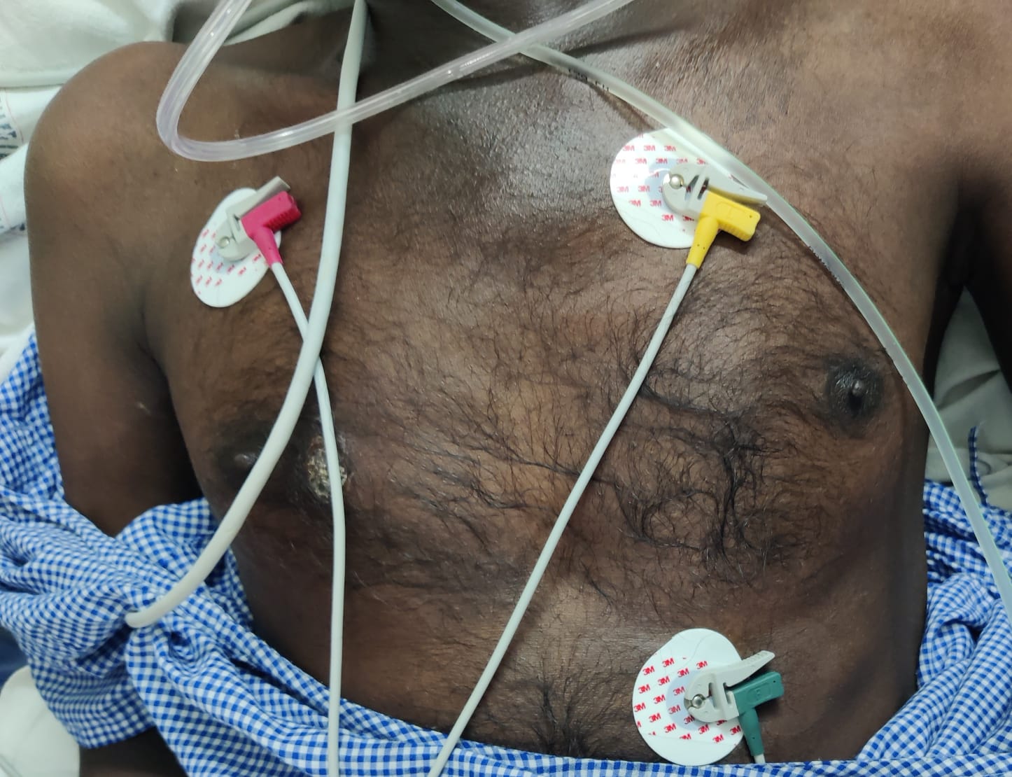
No scars, pulsation, engorged veins.
lesion present on beside right nipple
chest is bilaterally symmetrical
shape of chest - elliptical
bilateral airway entry present
trachea - Midline
Auscultation-
wheezing and Krebs heard diffusely around chest
Percussion- right left
supra clavicular resonant. resonant
infra clavicular resonant resonant
supra mammary resonant resonant
infra mammary resonant resonant
axillary resonant resonant
supra axillary resonant resonant
infra axillary resonant resonant
supra scapular resonant resonant
infra scapular resonant resonant
ABDOMINAL EXAMINATION
shape- scaphoid
tenderness no
no palpable mass
liver not palpable
spleen not palpable
CNS EXAMINATION
speech normal
no focal neurological deficits seen
INVESTIGATIONS
Complete blood picture
hemoglobin - 8.6 gm/dl
total count - 19,200cells/cumm
neutrophils - 91%
lymphocytes - 3%
pcv - 27.6%
blood group A+
interpretation- Normocytic normochromic anemia with neutrophilic leukocytosis
URINE EXAMINATION
albumin ++
sugar nil
pus cells 2-3
epithelial cells 2-3
Red blood cells 4-5
random blood sugar - 124 mg/dl
Renal functional test
urea 154/dl
creatinine 5.9mg/dl
uric acid 8.7 mg/dl
sodium 133mEq/L
Serum Iron- 74 ug/dl
Liver functional test
Alkaline phosphate 312 mg/dl
total protein 6.2 gm/dl
albumin 3.04gm/dl
ABG ANALYSIS
pH - 7.13
pCO2 - 34.1 mmHg
pO2 - 54.6 mmHg
HCO3 -11.1 mmol/L
O2 saturation 95.9%
GENERAL EXAMINATION FINDINGS
- Ryles feed -100ml milk +protein powder 2 scoops
- Neb. Budecort and duolin 8hrly
- Inj. piptaz 2.25 gm iv-TID
- Inj.Lasix 40mg IV/BD
- Inj.Pan 40mg IV/OD
- Inj.Hydrocort 100 mg IV/BD
- Tab.Telma H
- Dialysis
- strict I/O charting
- Monitor vitals
*For the past 6 months, the patient has been experiencing pain continuously every day, which did not resolve even on taking oral medication.
*Complains of weight loss around 15 kg in the past 6 months.
"In August 2022 ,the patient had an episode of abdominal pain and vomiting, for which he was admitted to a hospital. A CT of abdomen was performed and a lump was found in the pancreas. On further investigation, it was found that the lump was not cancerous. The patient was given symptomatic treatment and discharged when he was stable."
*From the past 6 months, the patient also complains of severe pain in both the legs ( in the calf region ), below the knee, which developed after trauma . The pain would start while sleeping or sitting for a long time. It is muscular in nature. The pain would get reduced by massaging the area. The pain is so severe that he is not able to sleep. He does not get the pain while walking. It is not associated with any changes in the overlying skin or swelling or muscle cramps.
*SMOKING- 2 packs a day from when he was in college. 1 pack a day from 6 months.
*ALLERGIES- no
Shape of the abdomen: normal
Umbilicus: normal
* No visible pulsations
* All quadrants of abdomen are moving equally on respiration.
* Grey turner sign ( bluish discolouration of flanks) and Cullens sign( bluish discolouration of periumbilical area ) are negative [ These are +ve in patients with severe pancreatitis with Haemorrhage ]
Palaption:
* No local rise of temperature
* Slight Tenderness present over left hypochondriac region.
* Guarding and rigidity : present
* No palpable masses found
* Liver and spleen are not palpable
Percussion :
* Liver span: normal
Ascultation:
* Sluggish bowel sounds are heard.
2) Respiratory system:
Slight left side deviation of the nasal septum that developed after trauma ,which did not affect his daily life
* Bilateral Normal vesicular breath sounds are heard.
*Position of trachea : central
3) CVS:
* S1 and S2 heart sounds are heard
*No murmurs
4) CNS:
* No focal neurological defecits
Provisional diagnosis:
Acute pancreatitis
INVESTIGATIONS:
* Imaging:
1) CE CT ( Contrast Enhanced CT):
- Get link
- X
- Other Apps


Comments
Post a Comment