long case
CHIEF COMPLAINTS
⬩ Altered gait since 2 year
⬩ Unable to walk since 3 months
⬩ Stiffness of all limbs since 15 days
⬩ decreased appetite since 15 days
⬩ weakness of all limbs since 15 days
⬩ unresponsiveness since 4 days
HISTORY OF PRESENTING ILLNESS
The patient was alright 2 years ago then he started developing generalised weakness, and complained that he felt like he was walking on pins few times. Generalised weakness caused him to stop working as a goldsmith which he has been doing since his youth. He would still do his own activities like bathing, eating, brushing etc. His routine before included him waking up at 5 am and carrying out his daily activities like brushing, bathing and eating his breakfast before going to work. He would work until 4 pm and then resume his other activities till sleep.
Now he would spend the majority of his time watching tv and carrying out his other routine activities except going to work.
1 year ago he started feeling intermittent weakness in his limbs and feeling pins and needles sensation on his feet. The weakness was gradual in onset and progressing and intermittent. He would go out even less now and spent majority of his time on the bed, uninterested to even watch tv. He was being taken to the hospital multiple times but with no proper diagnosis.
6 months ago he started being scared of walking as he was scared he would fall down, and the pricking sensation in the foot increased.
3 months ago he was taken to a hospital on one such episode of weakness, where they took a MRI in Nalgonda which showed:
- Hydrocephalus
- early Parkinson's changes
They advised to relieve the CSF pressure but due to financial reasons they did not go forward with the procedure.
20 days ago he developed typhoid fever which was diagnosed locally after which he deteriorated even more.
15 days ago he developed stiffness of all the limbs and also weakness in all the limbs. His wife massaged his legs with oil with which his symptoms relieved a little.
15 days ago he started eating less ( decreased appetite )
5 days ago he stopped feeding completely.
3 days ago he developed fever not associated with chills and rigours and became unconscious.
PAST HISTORY
Known case of hypertension since 5 years
Known case of diabetes since 3 years - taking glimipiride 2 mg + metformin 500 mg
No H/O epilepsy, asthma, tuberculosis, CAD
TREATMENT HISTORY
- antihypertensives ( drug unknown )
- oral hypoglycemics - glimipiride 2 mg + metformin 500 mg
- NSAIDS frequently
PERSONAL HISTORY
Diet: mixed
Appetite: decreased
Sleep: adequate
Bowel and bladder: normal
No allergies
Chronic alcoholic since he was 20 years of age and stopped 2 years back - he used to take 250 ml everyday
FAMILY HISTORY
No known family history
GENERAL EXAMINATION
The patient is conscious, not coherent, not cooperative, not oriented to time, place and person.
He is poorly built and malnourished.
VITALS
Temperature: afebrile
BP: 110/70 mm Hg
HR: 92 bpm
RR: 22 cpm
SpO2: 96 %
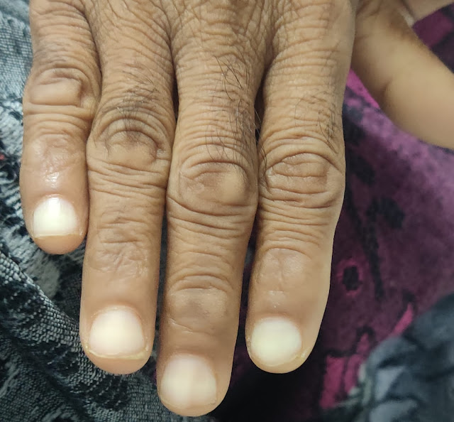
Pallor present
Skin is dry and patient looks dehydrated
Skin pinch test: skin goes back slower
No cyanosis, icterus, clubbing, lymphadenopathy, edema
SYSTEMIC EXAMINATION
CVS:
Inspection:
No dilated veins
Apex beat not visible
No pulsations seen
Palpation:
All inspectory findings were confirmed
Apex beat- diffuse
No palpable murmurs
Auscultation:
S1, S2 heard
No murmurs
Percussion: Normal
RESP:
Inspection:
Chest is symmetrical
Trachea central
Palpation:
All inspectory findings confirmed
Chest movements normal
Percussion:
Resonant note
Auscultation:
NVBS and BAE
PER ABDOMEN:
Scaphoid
Visible epigastric pulsations
No engorged veins/scars/sinuses
Soft, non tender
No organomegaly
Tympanic node heard all over the abdomen
Bowel sounds present
CENTRAL NERVOUS SYSTEM:
Higher mental function: conscious, non coherent, not oriented to time, place and person.
Language - APHASIC
Unable to write or read
GCS ( Glasgow coma scale)
Eye opening: to speech - 3
Verbal response: no response - 1
Motor response: flex to withdraw from pain - 4
Total score: 8
CRANIAL NERVE EXAMINATION:
CN 1: OLFACTORY NERVE - cannot be examined
CN 2: OPTIC NERVE - cannot be examined
CN 3: OCULOMOTOR NERVE - Normal
CN 4: TROCHLEAR NERVE - Normal
CN 5: TRIGEMINAL NERVE - Normal
CN 6: ABDUCENS NERVE - Normal
CN 7: FACIAL NERVE - Normal
CN 8: VESTIBULOCOCHLEAR NERVE- Normal
CN 9: GLOSSOPHARYNGEAL NERVE - gag reflex present
CN 10: VAGUS NERVE
CN 11: SPINAL ACCESSORY NERVE - cannot be examined
CN 12: HYPOGLOSSAL NERVE - cannot be examined
SENSORY SYSTEM: Cannot be examined
MOTOR SYSTEM: Muscle wasting present, no cramping, no involuntary movements.
BULK: Less
TONE: Hypertonicity in flexors and extensors of both upper and lower limbs
All the limbs were in flexed positing.
Lead pipe rigidity seen.
POWER:
Neck: cannot be elicited
3/5 in the upper limbs
Lower limbs power couldnt be elicited
REFLEXES:
SUPERFICIAL:
- Corneal reflex: present
- Conjunctival reflex: present
- Abdominal reflex: present
- Plantar reflex: present
- pharyngeal reflex: present
- Palatal reflex present
- Cremasteric reflex present
DEEP:
UPPER LIMB:

LOWER LIMB: Cannot be elicitedNo primitive reflexes
No meningeal signs
Gait cannot be seen
PROVISIONAL DIAGNOSIS
- Altered sensorium due to hypovolemic hyponatremia
- Parkinson’s disease
INVESTIGATIONS
Serum calcium: 8.3 mg/dl
Serum osmolality: 255 m OSM/kg
Sodium: 122 mEq/L
Potassium: 4.5 mEq/L
Calcium: 0.94
Blood urea: 53 ( 12-42 Normal)
RBS: 163 (100-160)
LFT:
AST: 47 IU/L
ALT: 50 IU/L
Alkaline phosphatase: 196 IU/L(56-119)
Total protein: 5.3 gm/dl
Albumin: 2.4 gm/dl
T3: 0/39 ng/ml
Spot urine protein: 4.0 mg/dl
Spot urine creatinine: 89 mg/dl
Ratio: 0.44
Serum creatinine: 0.7 mg/dl
Protocol:.
Axial TI, T2, FLAIR, DWI & SWI sequences, Coronal T2 and Sagittal TI sequences are taken.
OBSERVATIONS:
Disproportionate dilatation of lateral ventricle, third & fourth ventricle.
(Evan index 0.32).
Prominent sylvian fissure bilateral mild bowing of corpus collosum. Mild crowding of gyri at vertex.
Mild diffuse cerebral & cerebellar atrophy noted.
Bilateral confluent FLAIR hyperintense signal in bilateral periventricular white matter, corona radiata & centrum semiovale, central pons, MCP - Small vessel ischemic changes.
Mild atrophy of mid brain & pons.
Focal area of restricted diffusion seen in right superior frontal region with subtle low signal on ADC & FLAIR hyperintense signal - Suggestive of subacute infarct.
Micro hemorrhage in left thalamus, central pons, periventricular white matter.
Rest of the cerebral parenchyma shows normal gray/white matter differentiation.
Basal ganglia & thalami are normal.
Cerebellum normal.
Cranio-vertebral and Cervico-medullary junctions are normal.
Sella, pituitary and parasellar regions are normal.
Stalk and hypothalamus are normal. Posterior pituitary bright spot is normal.
Orbit and globe contents are normal.
IMPRESSION:
Focal area of restricted diffusion in right superior frontal region with subtle low signal & FLAIR hyperintense signal - Suggestive of subacute infarct.
Dilatation of ventricle with mild bowing of corpus collosum & crowding of gyri at vertex - Likely suggestive of normal procedure hydrocephalus.
Mild diffuse cerebral & cerebellar atrophy with moderate - severe small vessel ischemic changes.
Miero hemorrhage as described above - Likely hyperintensive microangiopathic
TREATMENT
- 3% NACL @ 15 ml/ hr
- Head end elevation upto 30 degrees
IV fluids:
- Ryles tube feeding with water and milk 100 ml
- Ringer lactate, Normal Saline
- Syndopa plus tablet 125 mg
----------------------------------------------------------------------------------------------------------------------------------------------------
short case
CHIEF COMPLAINTS
- Pain abdomen since 2 months
- Abdominal bloating since 2 months
- Blood in stools 2 months ago
- Shortness of breath on exertion since 1 month
- Palpitations since 1 month
HISTORY OF PRESENTING ILLNESS
The patient was apparently alright 2 months ago and then he developed pain in the right lumbar region which then became diffuse in nature. The pain was intermittent and had no aggravating or relieving factors. It was a dull aching type of pain. He also complained of bloating and belching with no H/O of any loose stools or vomitings. He then got himself checked in a local hospital where during the routine investigations it was found that he has low haemoglobin and he then underwent 2 blood transfusions and also used other syrups for it. Around that time after the blood transfusions he found out he had passed blood in his stools. He then developed shortness of breath on exertion since 1 month and palpitations since 1 month. He also gives a history of easy fatiguability and dizziness.
PAST HISTORY
H/O haemorrhoids 1 year ago.
No H/O DM, HTN, asthma, epilepsy, CAD
FAMILY HISTORY
No similar complaints in the family
PERSONAL HISTORY
DIET: Mixed
APPETITE: decreased
SLEEP: adequate
B&B: regular
He is a chronic alcoholic since 25 years (the last binge of alcohol was 2 months ago) He used to drink a type of Local alcohol and then changed to whisky.
No allergies
GENERAL EXAMINATION
The patient was conscious, coherent, cooperative, well oriented to time, place, person. He was moderately built and moderately nourished.
Pallor: Present
No icterus, cyanosis, clubbing, lymphadenopathy, pedal edema
VITALS:
Temperature: Afebrile
BP: 120/80 mm Hg
PR: 94 bpm
RR: 30 cpm
PER RECTAL EXAMINATION
No Skin tag at 6 o clock position and grade 1 haemorrhoids at 7 o clock position.
SYSTEMIC EXAMINATION
CVS - S1, S2 heard
RS: BAE +
P/A: soft, non tender
CNS: NAD
PROVISIONAL DIAGNOSIS:
Iron deficiency anemia secondary to Nutritional anemia/ chronic blood loss/ Haemorrhoids.
INVESTIGATIONS:
ULTRASOUND:
Test | Result |
HAEMOGLOBIN | # 3.1 gm/dl |
TOTAL COUNT | # 10,800 cells/cumm |
NEUTROPHILS | 72% |
LYMPHOCYTES | # 18 % |
EOSINOPHILS | 1% |
MONOCYTES | 9% |
BASOPHILS | 0% |
PCV | # 13.5 vol% |
M C V | # 54.9 fl |
M C H | # 12.6 pg |
MCHC | # 23.0 % |
RDW-CV | # 24.5 % |
RDW-SD | 46.9 fl |
RBC COUNT | # 2.46 millions/cumm |
Platelet count | 6.6 lakhs/cu.mm
|
Peripheral smear:
RBC: microcytic hypochromic with few pencil forms
Platelet count increased on smear
Blood group: O positive
Serum creatinine: 0.7 mg/dl
LFT:
Test | Result |
Total Bilurubin | 0.58 mg/dl |
Direct Bilurubin | # 0.24 mg/dl |
SGOT(AST) | 15 IU/L |
SGPT(ALT) | 10 IU/L |
ALKALINE PHOSPHATE | # 208 IU/L |
TOTAL PROTEINS | 6.8 gm/dl |
ALBUMIN | 3.7 gm/dl |
A/G RATIO | 1.24 |
TREATMENT:
1. 2 PRBC Transfusions done on 19th and 21st of November.
2. TAB. OROFER - XT
3. TAB. RANTAC
4. INJ. Ferric carboxymaltose 500 mg/ iv
TIMELINE:


Comments
Post a Comment