1801006146 CASE PRESENTATION
long case
CHIEF COMPLAINTS:
A 56 y/o female from Chityala, lemon seller by occupation, presented with chief complaints of:
→ Fever since 10 days
→ Abdominal pain since 7 days
→ Shortness of breath since 7 days
HISTORY OF PRESENTING ILLNESS:
Patient was apparently asymptomatic 10 days ago, then she developed fever which was of insidious onset, low grade, intermittent type, associated with chills and rigours, with no aggravating factors, and relieved on medication.
She also complains of cough with scanty white sputum for the first 2 days.
She had decreased urine output since 10 days.
She complains of abdominal pain since 7 days, situated in the right hypochondrium, which was mild during the 1st and 2nd day, and increased on the 3rd day. It is of pricking type, non-radiating, no aggravating factors and is relieved on medication.
She complains of shortness of breath since 7 days, which was insidious in onset, grade II; not associated with orthopnea and paroxysmal nocturnal dyspnea.
She complains of vomiting on the day of admission (13.03.2023), which was non-bilious, non-projectile, non foul smelling, non blood stained, and watery in consistency. She had an episode of vomiting before admission and one after admission.
PAST HISTORY:
3 moths ago: she developed itching over her left leg after which she slowly developed a swelling on the same leg, upto the knee. She went to a local hospital and was given intramuscular injection in her left gluteal region. The swelling subsided within 10 days and the injection site hardened. Later, she was given 2 injections at the same site for fever and abdominal pain. It gradually progressed in size and she was confined to bed due to pain. It developed into an abscess.
20 days ago: she developed fever, nausea, loss of appetite, and generalised weakness for which she went to a local RMP who gave her medication which relieved her symptoms.
18 days ago: she had abdominal pain for which she went to a local hospital and was diagnosed with acute kidney injury.
She is a known case of hypertension since 1 year and is taking Telma 40 mg.
No h/o diabetes, coronary artery disease, tuberculosis, asthma, epilepsy, thyroid disease.
PERSONAL HISTORY:
Diet: mixed
Appetite: decreased
Sleep: adequate
Bowel movements: regular
Bladder movements: decreased
Addictions: toddy occasionally since 15 years; alcohol everyday since 6 years
Allergies: none
FAMILY HISTORY:
No similar complaints in the family.
GENERAL EXAMINATION:
Patient was conscious, coherent, cooperative, moderately built and nourished, well oriented to time, place and person.
Pallor: present
Icterus: absent
Cyanosis: absent
Clubbing: absent
Lymphadenopathy: absent
Pedal edema: present
Vitals:
Temperature: afebrile
Pulse rate: 74 bpm
Respiratory rate: 18 cpm
Blood pressure: 130/80 mm Hg
SYSTEMIC EXAMINATION:
The patient was examined after consent was taken.
ABDOMEN:
Inspection:
Shape: round, slightly distended
Flanks: full
Umbilicus: inverted
No scars, no sinuses ,no dilated veins
Striae present
All quadrants moving equally with respiration
Right hypochondrial bulge seen
Palpation:
No local rise of temperature
Tenderness present in the right hypochondriac region
Liver: enlarged, surface smooth, rounded edges, firm in consistency, tender
No splenomegaly
Percussion:
Liver span of 14 cm, 4cm below the costal margin
Fluid thrill and shifting dullness absent
Auscultation:
Bowel sounds heard
No bruit heard
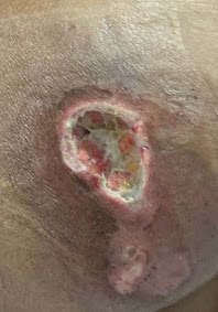
LOCAL EXAMINATION OF LEFT GLUTEAL REGION:
Wound size of 4×5 cm in left buttock, necrotic patch seen, induration seen, necrotic patch removed, abscess drained.
On inspection: 4×5 cm, margins are well defined ,edges are sloping and floor has slough and granulation tissue.
CVS examination:
S1, S2 heard; no murmurs
Respiratory system examination:
Inspection:
Shape of the chest : elliptical, b/l symmetrical
Both sides move equally with respiration
No scars, sinuses, engorged veins, pulsations
Palpation:
Trachea: central
Expansion of chest: symmetrical
Auscultation:
B/l air entry present
Normal vesicular breath sounds heard
CNS examination:
No neurological deficits
INVESTIGATIONS:
→ USG abdomen:
Findings: 5 mm calculus noted in gall bladder with GB sludge
Impression: cholelithiasis with GB sludge
Grade 2 fatty liver with hepatomegaly
→ Renal function tests:
15.03.2023:
Test | Result |
Blood urea | 64 mg/dL |
Serum creatinine | 1.6 mg/dL |
Serum Na | 125 mEq/L |
Serum K | 3.0 mEq/L |
Serum Cl | 88 mEq/L |
Test | Result |
Blood urea | 70 mg/dL |
Serum creatinine | 1.1 mg/dL |
Serum Na | 132 mEq/L |
Serum K | 3.2 mEq/L |
Serum Cl | 98 mEq/L |
Test | Result |
Blood urea | 60 mg/dL |
Serum creatinine | 1.1 mg/dL |
Serum Na | 133 mEq/L |
Serum K | 3.6 mEq/L |
Serum Cl | 99 mEq/L |
→ Complete urine examination:
Colour: pale yellow
Appearance- clear
Specific gravity: 1.010
Albumin: trace
Pus cells: 2 - 4/hpf
Epithelial cells: 2 - 3/hpf
Sugar: nil
→ X-ray abdomen:
→ Hemogram:
Test | Result |
Hemoglobin | 9.6 g/dL |
Total count | 15500 cells/cu mm |
Neutrophils | 75% |
PCV | 29.6 vol% |
RBC count | 3.1 million/cu mm |
→ Liver function tests
Test | Result |
Total bilirubin | 2.6 mg/dL |
Direct bilirubin | 1.1 mg/dL |
Indirect bilirubin | 1.5 mg/dL |
Alkaline phosphatase | 193 IU |
AST | 37 IU |
ALT | 21 IU |
Total protein | 7 g/dL |
Albumin | 4.3 g/dL |
Globulin | 2.7 g/dL |
A:G ratio | 1.6 |
PROVISIONAL DIAGNOSIS:
Acute cholecystitis with hepatomegaly
Acute kidney injury
Gluteal abscess
TREATMENT:
→ Liquid diet
→ Inj. PAN 40 mg i.v./OD
→ Inj. PIPTAZ 2.25 mg/i.v./TID
→ Inj. METROGYL 500 mg/i.v./TID
→ Inj. Zofer 4 mg i.v./SOS
→ Inj. NEOMOL 1 gm i.v./SOS
→ T. Paracetamol 650 mg p.o./TID
→ T. CINOD 10 mg p.o./OD
→ I.v. fluids 1 unit NS, RL, DNS 100 ml/hr
→ Inj. Buscopan 10 mg OD
→ Pneumatic compressor bed
→ 2-hourly change in position
----------------------------------------------------------------------------------------------------------------------------------------------------
short case


Comments
Post a Comment